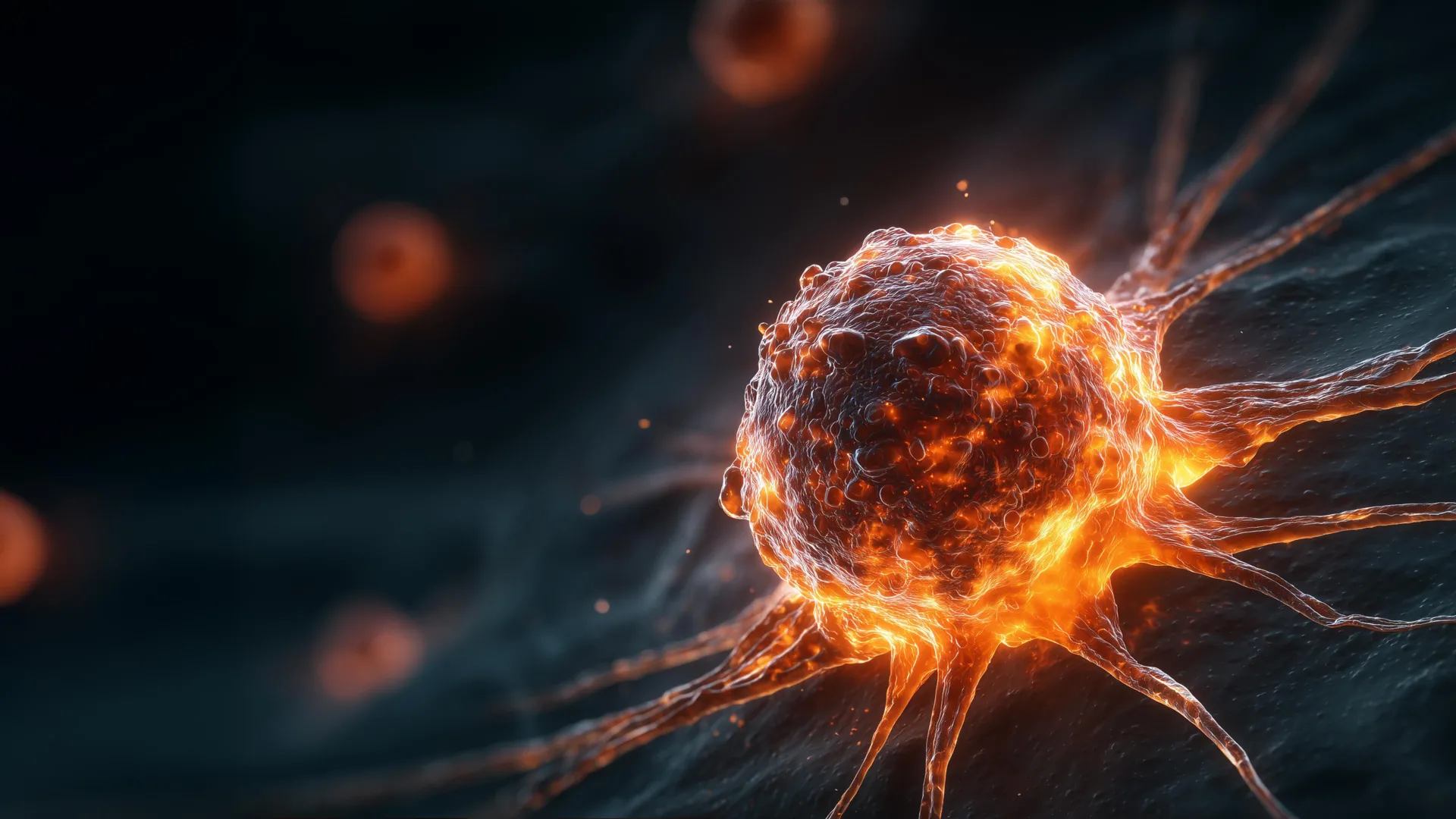
Unlocking Ferroptosis: A Step-by-Step Guide to Counteracting Vitamin B2's Shield in Cancer Cells
Overview
Recent research has revealed a surprising paradox: vitamin B2 (riboflavin), an essential nutrient, may actually help cancer cells survive by protecting them from a form of programmed cell death known as ferroptosis. This guide explains the science behind this discovery and provides a detailed laboratory protocol for using a riboflavin analog, roseoflavin, to dismantle that protective shield and trigger cancer cell death. By the end, you'll understand the molecular mechanisms and have a clear experimental path to reproduce these findings.
Why This Matters
Ferroptosis is a regulated cell death process driven by iron-dependent lipid peroxidation. Cancers often rely on antioxidants to evade ferroptosis. Vitamin B2 serves as a precursor for flavin adenine dinucleotide (FAD), a cofactor for glutathione reductase (GSR). GSR regenerates reduced glutathione (GSH), a key antioxidant that neutralizes lipid peroxides. Thus, riboflavin indirectly strengthens the cancer cell's defense against ferroptotic death. Roseoflavin, a natural riboflavin analog, competes with riboflavin for incorporation into FAD, leading to dysfunctional GSR and rendering cells susceptible to ferroptosis.
Prerequisites
Background Knowledge
- Basic cell culture techniques (sterile technique, media preparation, passaging)
- Understanding of reactive oxygen species (ROS) and lipid peroxidation
- Familiarity with cell viability assays (MTT, CellTiter-Glo) and ferroptosis detection (e.g., C11-BODIPY staining)
Materials & Reagents
- Cancer cell lines (e.g., HT-1080, MDA-MB-231) – use mycoplasma-free cultures
- Roseoflavin (CAS 51093-55-2) – dissolve in DMSO at 10 mM stock
- Riboflavin (vitamin B2) – prepare in culture medium at desired concentrations
- Ferroptosis inducer: erastin (10 µM) or RSL3 (1 µM) – optional positive control
- Ferroptosis inhibitor: ferrostatin-1 (1 µM) or liproxstatin-1 – to confirm specificity
- Cell culture medium (DMEM or RPMI with 10% FBS, penicillin/streptomycin)
- Lipid peroxidation sensor: C11-BODIPY 581/591 (Thermo Fisher)
- Glutathione assay kit (e.g., GSH/GSSG-Glo assay)
- GPX4 antibody (for Western blot) – to monitor protein levels
Step-by-Step Instructions
1. Cell Culture Preparation
Seed cancer cells in 96-well plates (for viability) or 6-well plates (for biochemical assays) at 5,000 cells per well in complete medium. Allow attachment overnight at 37°C, 5% CO2. Ensure cells are in logarithmic growth phase.
2. Dose-Response Curve for Roseoflavin
Prepare serial dilutions of roseoflavin in culture medium (e.g., 0, 5, 10, 20, 50, 100 µM). Add to wells in triplicate. Incubate for 24–48 hours. Measure cell viability using an MTT or luminescent ATP assay. Determine IC50 values. Typically, cancer cells show reduced viability at ≥20 µM roseoflavin when combined with low riboflavin conditions.
3. Simulate Vitamin B2 Protection
In parallel, supplement culture medium with 1–10 µM riboflavin (physiological range). Repeat roseoflavin treatment. Observe that riboflavin rescues cells from roseoflavin toxicity, confirming the competitive mechanism.
4. Quantify Ferroptosis Markers
After 24-hour treatment with roseoflavin (choose a sub-IC50 dose, e.g., 10 µM), harvest cells for analysis:
- Lipid peroxidation: Incubate cells with C11-BODIPY (2 µM, 30 min). Analyze by flow cytometry or fluorescence microscopy. Increased oxidation shifts fluorescence from red to green.
- GSH/GSSG ratio: Use a commercial kit. Roseoflavin-treated cells should show decreased GSH and increased GSSG, indicating oxidative stress.
- GPX4 expression: Perform Western blot. Expect reduced GPX4 levels if cell death is ferroptotic, though acute effects may be post-translational.
5. Confirm Ferroptosis Specificity
Co-treat cells with roseoflavin and ferrostatin-1 (1 µM) or liproxstatin-1 (1 µM). If cell death is rescued, ferroptosis is the primary mode. Compare with necroptosis or apoptosis inhibitors (e.g., Nec-1, Z-VAD-FMK) to rule out other pathways.
6. Assess Riboflavin Uptake and FAD Levels
Measure intracellular FAD by HPLC or using a fluorescence-based assay. Roseoflavin reduces FAD levels by competing for riboflavin kinase (RFK) and FAD synthetase. This step links the molecular mechanism to the observed phenotype.
7. Optional: In Vivo Validation
Using a xenograft mouse model, administer roseoflavin (i.p. injection, 25 mg/kg daily) with or without dietary riboflavin restriction. Monitor tumor growth and harvest tumors for ferroptosis markers. Ensure ethical approval before proceeding.
Common Mistakes
Overlooking Riboflavin in Culture Medium
Standard DMEM contains ~1 mg/L (2.66 µM) riboflavin. This basal level can mask roseoflavin effects. Use riboflavin-free medium for critical experiments, or include a riboflavin-depleted serum (dialyzed FBS). Without controlling this variable, results may be inconsistent.
Using Too High Roseoflavin Concentrations
Roseoflavin at >100 µM can cause off-target effects (e.g., mitochondrial toxicity). Always include a dose-response and verify specificity with genetic modulation (e.g., RFK knockdown) if possible.
Misinterpreting Viability Data
Ferroptosis is often slower than apoptosis. Measure viability at multiple time points (24, 48, 72 hours). A single endpoint can miss delayed effects. Also, combine viability with lipid peroxidation to confirm mechanism.
Ignoring Cell Line Variability
Not all cancer cell lines respond equally. Mesenchymal-type cancers (e.g., HT-1080) are more sensitive to ferroptosis than epithelial types. Test multiple lines to generalize findings.
Forgetting Negative Controls
Always include vehicle (DMSO) controls at equivalent concentrations. Roseoflavin is light-sensitive; protect solutions from light during storage and handling. Also, verify that your ferroptosis inhibitors are active (e.g., use erastin as positive control).
Summary
This guide details the experimental approach to unraveling the vitamin B2–ferroptosis paradox. By using roseoflavin to competitively inhibit riboflavin utilization, researchers can overcome the cellular shield that protects cancer cells from ferroptotic death. The protocol covers cell culture, drug treatment, key assays (lipid peroxidation, GSH levels, GPX4 status), and specificity testing. Common pitfalls include media riboflavin content and dose selection. Mastering this protocol will empower scientists to explore ferroptosis-based therapies and understand how nutritional factors influence cancer survival.